Applications
Here we highight a few applications of our system. More applications are described in detail in other publications (see menu bar on the left).
Cone-beam CT with flat-panel detectors
The recent emergence of various types of flat-panel x-ray detectors and C-arm gantries now enables the reconstruction of novel imaging platforms for a wide variety of clinical applications. Many of these applications require interactive 3D image generation, which cannot be satisfied with inexpensive PC-based solutions using the CPU. We present a solution based on commodity graphics hardware (GPUs) to provide these capabilities. While GPUs have been employed for CT reconstruction before, our approach provides significant speedups by exploiting the various built-in hardwired graphics pipeline components for the most expensive CT reconstruction task, backprojection. We show that the timings so achieved are superior to those obtained when using the GPU merely as a multi-processor, without a drop in reconstruction quality. In addition, we also show how the data flow across the graphics pipeline can be optimized, by balancing the load among the pipeline components. The result is a novel streaming CT framework that conceptualizes the reconstruction process as a steady flowof data across a computing pipeline, updating the reconstruction result immediately after the projections have been acquired. Using a single PC equipped with a single high-end commodity graphics board (the Nvidia 8800 GTX), our system is able to process clinically-sized projection data at speeds meeting and exceeding the typical flat-panel detector data production rates, enabling throughput rates of 40–50 projections/s for the reconstruction of 5123 volumes.
For further detail see the paper: Xu / Mueller, "Real-Time 3D Computed Tomographic Reconstruction Using Commodity Graphics Hardware," Physics in Medicine and Biology, 52:3405–3419, 2007.
Ultrasound 3D breast mammography: Tomographic reconstruction in refractive media
Transmission Ultrasound Computed Tomography (UCT) is strongly affected by the acoustic refraction properties of the imaged tissue, and proper modeling and correction of these effects is crucial to achieving high-quality image reconstructions. In earlier work, we derived a method that can account for these refractive effects by solving the governing Eikonal equation within an iterative reconstruction framework using a wave-front tracking approach. Excellent results can be obtained, but at considerable computational expense. To overcome these obstacles, we accelerated three Eikonal solvers: the Fast Marching Method (FMM), the Fast Sweeping Method (FSM), and the Fast Iterative Method (FIM) on the NVIDIA Tesla multi-processor board, within our refractive Transmission Ultrasound CT framework. Using our accelerated FIM we were able to reconstruct breast phantoms of size 2562×44 (512 transducers in each of the 44 ring detectors) in less than 5 minutes. This is a time that can easily satisfy real-life clinical scenarios.meeting the interactive demands of clinical practice, without a loss in reconstruction quality.
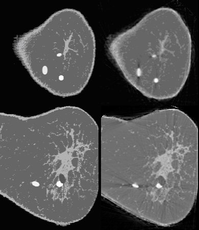
The left column shows two slices of an acoustic breast phantom derived from the Visible Female dataset and the right columns shows their UCT reconstructions (first row: slice close to the center, second row: slice close to the bottom).
For further detail see the paper: Li / Mueller et al., "Physical-Space Refraction-Corrected Transmission Ultrasound Computed Tomography Made Computationally Practical," MICCAI '08, (to be presented), September, 2008.